Schematic Image Of Cheek Cell Cbse Class 9 Science Practical
Cheek cell diagram Cheek cell bacteria cells human membrane nucleus using picture bacterial been single prokaryotic solved determine Cheek cells under microscope labeled
Solved Using this table from the Size Estimation module, | Chegg.com
Cheek cell diagram Diagram of cheek cells Cheek cells 400x stained
Solved using this table from the size estimation module,
Drawing cell cheek labelled human biological parts followed rules basic must there when some exportedHuman cheek cell lab report introduction Human epidermal cells diagramHow would you take the sample to prepare temporary stained mount of.
To prepare stained temporary mounts of human cheek cellSbi3u Cheek cell diagram5. describe an activity to observe cheek cells under a microscope..

Solved using this table from the size estimation module,
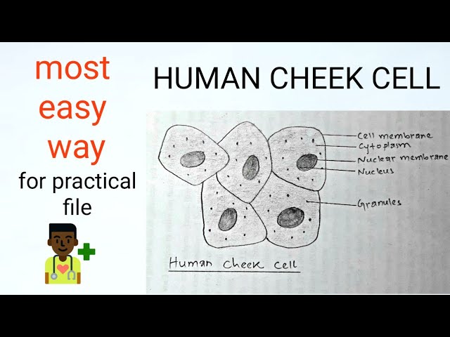
Cheek cell human draw labelling correctQuestions and answers on labeled/unlebled diagrams of a human cell Squamous epithelial cheek cells labeledCbse class 9 science practical skills – slide of onion peel and cheek cells.
Cheek onion cell vs cells comparing contrastingDraw the picture of cheek cells andlare ita Human cheek cell dna extractionCheek cells conclusion.

Draw the human cheek cell with correct labelling
Dic image of a cheek cellDraw cheek cell Cell cheek cells 400x stained human animal slide lab staticflickr picture c1 flickrDraw the human cheek cell with correct labelling.
Cells cheek microscope human under cell animal membrane do epitheliumHuman cheek cells under the microscope Cheek cell human stained temporary cells mounts prepare epithelial lab results layer work discussion studyCheek cell image using brightfield and darkfield microscopy. (a.

Top 197 + animal cheek cell
Cheek cell size cells human using 40x objective single module estimation table lens field organelle well solved determine writeHow to draw cheek cell step by step for beginners Darkfield microscopy brightfield cheek condenserSchematic image of a cheek cell.
Cheek cells under a microscopeImage result for human cheek cell diagram Cheek labelling ppz brainliestCheek dna extraction chromosomes mugeek vidalondon genetic.

Cheek dn labeled
Cheek cell labeled diagramCheek cells under microscope labeled .
.


Cheek cell image using brightfield and darkfield microscopy. (a

Cheek Cell Diagram

5. Describe an activity to observe cheek cells under a microscope.

Solved Using this table from the Size Estimation module, | Chegg.com
Squamous Epithelial Cheek Cells Labeled
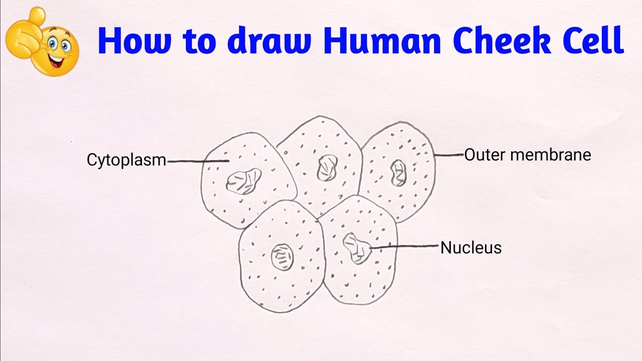
Cheek Cell Labeled Diagram

Cheek cells conclusion - Isolation and staining of cheek cells with